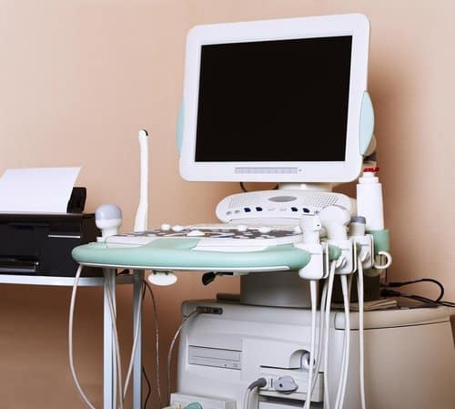
Duplex Venous Ultrasound

In a healthy leg, the veins push the blood upward in route to your heart. In individuals with venous insufficiency, quantities of blood can pool in the veins and cause a wide variety of medical symptoms including the presentation of varicose veins. Ultrasounds are more affordable and quicker to perform than other types of imaging scans like CT scans or MRIs however the outcome of the results rely heavily on the skills of the operator. Sometimes you may not want to trust the results of your ultrasound. For more detailed information on ultrasounds and what you need to know to make an informed decision, make sure to read the section below.
How do I get ready?
Don loose-fitting, comfy attire. It might be necessary to take off all of your clothes and valuables in the region being checked.
For the procedure, you might need to change into a gown.
Only if you’re having your abdominal veins examined is a fasting time required. In this situation, you’ll generally be told to wait six to eight hours before consuming any food or liquids besides water. A venous ultrasonography requires no additional specific preparation.
What to Expect During a Venous Reflux Exam
Duplex venous ultrasounds can be performed by a technician without the need for anesthesia. A handheld ultrasound “wand” is run over the length of the legs sending sound waves in to the leg. Those sound waves are reflected back (like a sonar or fish finder). The data is processed and used to create a series of images. These images allow the physician to examine any abnormalities in the veins and valves. It also shows the flow rate and direction of the blood in the veins.
These painless tests typically start with laying on a table. After applying a conductive gel, the vascular technologist will pass the wand over the area to be tested. Once the supine (lying down) assessment of your veins is performed, you will be asked to stand up to evaluate reflux. This is an essential step since gravity plays a significant role in venous insufficiency. The next part of the exam is crucial. The ultrasound tech squeezes and then releases your calf to see how this affects blood flow. The tech can use his or her hand or an automatic clamp. If you are suffering from venous insufficiency, this procedure will provoke the blood in your leg to travel back towards your foot. This can easily be detected by ultrasound. A duplex venous ultrasound exam generally takes between 30 minutes to an hour depending on if both legs are being evaluated or not.
Duplex venous ultrasounds are typically conducted as part of an overall varicose vein evaluation. Venous reflux examinations are non-invasive tests that don’t require a recovery period and are completely painless. No downtime or aftercare are required before returning to your normal daily activities. As no anesthesia is required, you can drive yourself home after the appointment is you normally drive yourself. I can be difficult to remove the ultrasound gel without a shower.
Are there any potential risks or side effects associated with a duplex venous ultrasound?
Duplex venous ultrasound is generally considered a safe and non-invasive procedure with minimal risks or side effects. However, as with any medical procedure, there are a few considerations to keep in mind:
- Discomfort: During the ultrasound, the technician may need to apply slight pressure to obtain clear images. This pressure can sometimes cause mild discomfort, especially if you have tender or sensitive areas.
- Rare allergic reactions: In some rare cases, individuals may have an allergic reaction to the gel used during the procedure. If you have a known allergy to ultrasound gel or similar substances, it is important to inform the healthcare provider beforehand.
- False-positive or false-negative results: Although duplex venous ultrasound is highly accurate, there is a slight possibility of false-positive or false-negative results. Factors such as operator technique, body habitus, or technical limitations of the equipment used can contribute to potential inaccuracies.
- Rare complications: While extremely rare, there is a slight risk of complications such as skin irritation or bruising at the site of gel application. These complications typically resolve on their own without requiring any specific treatment.
Can duplex venous ultrasound be used to monitor the progression of venous diseases?
Yes, duplex venous ultrasound can be effectively used to monitor the progression of venous diseases. Here are several points outlining its utility in this context:
- Baseline Assessment: Duplex venous ultrasound provides a baseline assessment of venous anatomy and function, establishing a starting point for monitoring disease progression.
- Quantitative Measurements: It allows for quantitative measurements of vein diameter, blood flow velocities, and valve function, which can indicate changes over time.
- Tracking Changes: By comparing sequential ultrasound examinations, healthcare providers can track changes in vein morphology, such as the development of varicose veins or the progression of chronic venous insufficiency (CVI).
- Monitoring Treatment Response: It helps assess the effectiveness of treatments like compression therapy, sclerotherapy, or surgical interventions by evaluating improvements or regressions in venous function and anatomy.
- Detection of Complications: Duplex ultrasound can detect complications such as recurrent deep vein thrombosis (DVT), thrombophlebitis, or venous ulcers, which may indicate disease progression.
Can duplex venous ultrasound detect abnormalities in perforator veins?
Yes, duplex venous ultrasound can detect abnormalities in perforator veins. Here are several points explaining its utility in this context:
- Localization of Perforator Veins: Duplex ultrasound accurately identifies the location and course of perforator veins, which connect superficial veins to deep veins.
- Assessment of Perforator Function: It evaluates the function of perforator veins by assessing blood flow velocities and detecting reflux (backward flow of blood) or obstruction.
- Mapping for Treatment Planning: It provides detailed mapping of perforator veins, essential for planning treatments such as sclerotherapy or surgical interventions to target specific abnormalities.
- Evaluation of Disease Severity: Duplex ultrasound helps assess the severity of venous diseases by identifying incompetent perforator veins that contribute to venous hypertension and complications like ulcers.
How soon will I receive the results of the duplex venous ultrasound?
The timing of receiving the results of a duplex venous ultrasound can vary depending on several factors, including the healthcare facility’s protocols and the urgency of your situation. In many cases, the ultrasound images are interpreted by a radiologist, who generates a report. This report is then typically shared with your referring healthcare provider.
In general, you can expect to discuss the results of the ultrasound during a follow-up appointment with your healthcare provider. The specific timing of this appointment will depend on the urgency of your condition and the availability of appointments.
